|
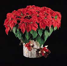
Fig. 1. Poinsettia: The Christmas flower in bloom (click image for larger view). |
|
D. Michael Benson
(mike_benson@ncsu.edu)
North Carolina State University, Raleigh;
Janet L. Hall
(jhall@eckeranch.com)
Paul Ecke Ranch, Encinitas, CA;
Gary W. Moorman
(gmoorman@psu.edu)
Pennsylvania State University, University Park;
Margery L. Daughtrey
(mld9@cornell.edu)
Cornell University, Long Island Hort Res Lab,
Riverhead, NY;
Ann R. Chase
(mtaukum@aol.com)
Chase Research Gardens Inc, Mount Aukum, CA;
Kurt H. Lamour
(lamourku@pilot.msu.edu)
Michigan State University, East Lansing
Benson, D.M., Hall, J.L., Moorman, G.W., Daughtrey, M.L., Chase, A.R. and Lamour, K.H. 2001. Poinsettia: The Christmas Flower. APSnet Features. Online. doi: 10.1094/APSnetFeature-2001-1201
History of the Poinsettia
The poinsettia, Euphorbia pulcherrima Willd., is a member of the family Euphorbiaceae. The genus Euphorbia contains some 700 to 1,000 species. It is characterized by a single female flower, without petals and usually without sepals, surrounded by individual male flowers all enclosed in a cup-shaped structure called a cyathium. The showy red, pink, white, or bicolored portion of the plant, popularly referred to as the flower, consists of modified leaves or bracts (Fig. 1).
The poinsettia is a native plant of Mexico and originated in a rather limited region near present day Taxco. Long before the arrival of Europeans, the Aztecs of central Mexico cultivated the plant and called it Cuetlaxochitl. Because of its brilliant color, the poinsettia was a symbol of purity to the Indians. It was highly prized by both King Netzahualcoyotl and Montezuma, but because of the high altitude climate, the plant could not be grown in their capital, now known as Mexico City. The Indians used poinsettia bracts to make a reddish-purple dye. They also made a medicine for fever from the plant’s latex.
During the 17th Century, a group of Franciscan priests settled near Taxco. They began to use the poinsettia in the Fiesta of Santa Pesebre, a native procession. Juan Balme, a botanist of the same period, mentioned the poinsettia plant in his writings. He described it as having large green leaves and a small flower surrounded by bracts, almost as if for protection. The bracts, he said, turned a brilliant red. Balme also found the plant flourishing on the slopes and in the valleys near Cuernavaca.

Fig. 2. Joel Roberts Poinsett (1779-1851) (click image for larger view). |
|
Poinsettias were first introduced in the United States in 1825 by Joel Roberts Poinsett (20,24) (Fig. 2). While serving as the first United States Ambassador to Mexico, he visited Taxco and found the flowers growing on the adjacent hillsides. Poinsett, a botanist of great ability, had some plants sent to his home in Greenville, South Carolina. They did well in his greenhouse and he distributed plants to botanical gardens and to horticultural friends, including John Bartram of Philadelphia. Bartram, in turn, supplied the plant to Robert Buist, a nurseryman who first sold the plant as Euphorbia pulcherrima, Willd. The name poinsettia, however, has remained the accepted name in English-speaking countries.
The modern era of poinsettia culture began with the introduction of the seedling cultivar Oak Leaf (Fig. 3). This cultivar was reported to have been grown originally in Jersey City, New Jersey, by a Mrs. Enteman in 1923. From 1923 until the early 1960s, all of the principal cultivars of commercial importance were selections or sports from this original Oak Leaf seedling.
|
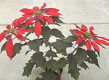
Fig. 3. Seedling cultivar ‘Oak Leaf,’ the progenitor of modern poinsettia cultivars (click image for larger view).
|
|
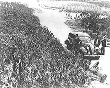
Fig. 4. Field production of poinsettia stock plants in Southern California in the mid-1930s (click image for larger view).
|
|
During the middle 1950s, poinsettia breeding programs were initiated at several institutions, including the Pennsylvania State University, the University of Maryland, the USDA Research Center at Beltsville, Maryland, and by a number of commercial horticulture firms including Azalealand in Lincoln, Nebraska; Paul Ecke Ranch in Encinitas, California (Fig. 4); Mikkelsen’s in Ashtabula, Ohio; Earl J. Small of Pinnellas Park, Florida; Yoder Brothers in Barberton, Ohio; Zieger Brothers in Hamburg, Germany; and Thormod Hegg & Son in Reistad, Norway. Dr. Robert N. Stewart, of the Agricultural Research Service of Beltsville, Maryland, used his genetic training to segregate desirable characteristics such as stiff stems, larger bracts, new colors, and lasting qualities. He contributed much in determining the character of mutation forms in poinsettias, and his cooperative efforts have been extremely helpful to the commercial hybridizers.
With the introduction of the cultivar Paul Mikkelsen in 1963 (Fig. 5), poinsettias entered a new era. This cultivar, with stiff stems and foliage retention characteristics, provided the trade with the first longer-lasting cultivar of commercial importance. Annette Heggä Red was introduced in Norway in 1964. This cultivar was quickly followed by a number of sports. The Hegg cultivars introduced an entirely new type of multi-flowered plant to the trade because of their ability to produce from five to eight blooms from a pinch, and because of their ease of production.
|
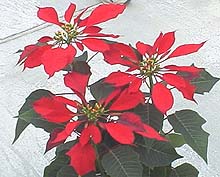
Fig. 5. Paul Mikkelsen poinsettia (click image for larger view). |
|
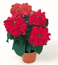
Fig. 6. Winter Rose the first cultivar with incurved bracts and foliage (click image for larger view).
|
|
In 1988 Eckespoint® Lilo was introduced. This was the of the first dark leaf poinsettia cultivars that were early flowering, recovered quickly after unsleeving, and had excellent foliage retention for the consumer. This cultivar required certain cultural techniques to insure good branching. In 1992, Eckespoint® Freedom™ was introduced. Eckespoint® Freedom™ contained the best characteristics of Eckespoint® Lilo while branching more consistently for the producers. Today there are over 100 poinsettia cultivars grown commercially, with one cultivar, Eckespoint® Freedom™ representing over 50% of the red market worldwide and 70-75% of that market consisting of poinsettias with red bracts. One final revolution to poinsettia cultivars was the introduction of Eckespoint® Winter Rose™ Dark Red in 1998 (Fig. 6). This cultivar was the first introduction in the “curly” family with dark red incurved bracts and deep dark green incurved foliage. (Adapted from Ecke, et al., 1990. The Poinsettia Manual [20].)
Today, poinsettias may be found in many different colors (Fig. 7) as well as product forms from mini poinsettias to large specimen trees and every size in between. Testifying to its success and popularity, the poinsettia is not only the most popular holiday flower, it is the number one flowering potted plant in the United States, with over 65 million plants sold nationwide in 2000 (61).
|
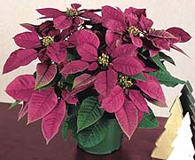
Fig. 7. Plum Pudding, the first poinsettia with purple bracts (click image for larger view). |
|
Diseases
Several diseases affect production of poinsettia, including foliar diseases such as Botrytis gray mold, powdery mildew, Alternaria blight, Xanthomonas blight, Erwinia blight, Phytophthora blight, and root diseases such as Pythium, Phytophthora and Rhizoctonia root rot. Powdery mildew is a fairly recent disease problem in poinsettia production that can develop explosively late in the crop production cycle. Scab caused by Sphaceloma poinsettiae, normally a disease problem only in states like Florida and Hawaii with subtropical climates, has been introduced nationwide the past couple of seasons with infected rooted cuttings from propagators in Central America. On the other hand, black root rot caused by Thielaviopsis basicola, a soilborne pathogen, was a serious disease of poinsettia in the 1950s and early 60s until the floriculture industry moved to soilless potting mixes. Since the 1920s poinsettias in the industry have exhibited a free-branching morphotype so that cultivars develop multiple branches from a single pinch resulting in many blooms. Recently, a phytoplasma etiology was described that explained the free branching habit (47,48).
Foliar Diseases
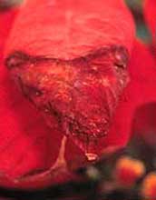
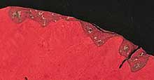
Gray mold. Botrytis cinerea (teleomorph Botryotinia fuckeliana (de Bary) Whetz.) can cause brown spots to form on poinsettia flower bracts (Fig. 8) and leaves. The bract damage is easily confused with bract burn (Fig. 9) caused by calcium deficiency, an imbalance among potassium, calcium, and magnesium or by an ammonium-calcium antagonism. The fungus readily invades tissue damaged in any way, including that caused by bract burn. After the true flowers have fully developed, Botrytis attacks and covers them with the typical smoky, gray spores. Poinsettias are particularly susceptible to gray mold late in the season when it can cause significant damage to the aesthetic quality of plants. Gray mold development also occurs during crop shipment if infections have occurred in the production greenhouse. Large, light brown to tan, slightly sunken cankers (Fig. 10) form when older stems are invaded through wounds on large branches or through cracked branch crotches. Defoliation and death of branches occur above cankers that girdle stems.
|
 |
|
 |
|
|
Fig. 8. Brown spots on poinsettia bracts caused by Botrytis (click image for larger view).
|
|
Fig. 9. Bract burn due to calcium deficiency (click image for larger view).
|
|
| |
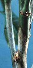
Fig. 10. Stem cankers caused by Botrytis infection (click image for larger view). |
|
Botrytis cinerea produces massive numbers of spores that are readily detached by air movement or rapid changes in relative humidity within the crop (39). Depending upon the plant infected, 104 to 107 spores/cm2 have been observed on leaf tissue (37). Peak spore concentrations in greenhouses can occur at midmorning and midafternoon (33,34). Spores can also be dispersed by splashing (40), in addition to wind dispersal. Sprinkler irrigation splashes spores from place to place and provides the moisture they need in order to germinate and invade plant surfaces. Although Botrytis thrives most under cool temperatures (20 to 25°C), it can in fact survive, sporulate, and continue to cause disease between 4 and 30°C (41).
It is important to manage Botrytis in the entire greenhouse because many different crops are susceptible to gray mold (37). If geraniums, primulas, impatiens, or other highly susceptible crops are grown in the same greenhouse as poinsettias, the fungus may form large numbers of spores on those very susceptible species and spread to the poinsettias. In general, fading flower tissue of almost any crop becomes a food source for Botrytis. In order to inhibit Botrytis development and minimize gray mold damage, the maintenance of low relative humidity (below 93%) within the crop canopy is crucial. Plants should be spaced and heating, venting, and air circulation regimes adjusted to can keep the humidity low throughout the poinsettia production season. This usually means that pots must be separated once or twice during production so that the canopy does not close. Irrigation systems that do not apply moisture to the above ground portions of the plants help to maintain an environment inhibitory to Botrytis growth. By maintaining low humidity, the need for fungicides is greatly reduced or eliminated. In operations where humidity can be consistently minimized within the canopy, fungicides are not needed.
Because the fungus readily attacks damaged, fading, or dead tissue, it is important to avoid damaging plants and, when possible, to remove damaged tissues. All plant debris should be promptly removed from the greenhouse or disposed of in covered containers to prevent the spores formed on this material from being dispersed to the crop. If these practices are followed, then fungicides can help in management. Fungicides including chlorothalonil, fenhexamid, fludioxonil, and copper and copper plus mancozeb can be used to protect foliage from Botrytis. Great care must be exercised in selecting and applying fungicides to bracts because phytotoxicity, in the form of yellow spots or bleaching of the bract color, can be caused by some materials. Furthermore, some fungicides leave a very visible residue that is unacceptable, particularly on darkly colored foliage and bracts. Botrytis populations in most greenhouses in the U.S. are resistant to the benzimidazole class of fungicides (thiophanate methyl). Some populations have multiple resistance to benzimidazoles and dicarboximides (iprodione and vinclozolin) (23,46,52,53,60) and other classes of fungicides must be employed.
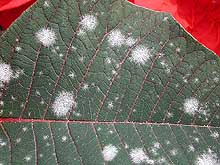
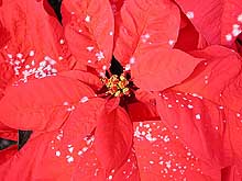
Powdery mildew. A powdery mildew disease on poinsettias was first seen in Mexico and Puerto Rico in the late 1980s, and in Pennsylvania and the Pacific Northwest in 1990. Over a hundred growers across the U.S. were affected by powdery mildew in 1992 (16). Since that time, the disease has appeared sporadically in North American greenhouses and has been seen in rare instances in Europe. Since there were no previous problems with powdery mildew in North America, many of the ornamental fungicides used to control powdery mildew on other plants were not at the time labeled to allow application to poinsettias. Growers were extremely concerned that application of fungicides to poinsettias late in the growing season would be injurious to the bracts. There was also a tendency at first to underestimate the impact that powdery mildew would have on the poinsettia crop. Not only did white colonies mar the leaves (Fig. 11), but the fungus formed dramatic white colonies on colored bracts, rendering plants unsalable (Fig. 12).
 |
|
 |
|
Fig. 11. Colonies of powdery mildew on a leaf (click image for larger view). |
|
Fig. 12. Colonies of powdery mildew on flower bracts (click image for larger view). |
The pathogen is an Oidium sp. (only the anamorph stage has been observed). It has no known hosts other than the poinsettia, which means that unless growers keep poinsettias over in the greenhouse from year to year, it is not likely to survive except in frost-free climates where poinsettias live year-round as landscape ornamentals. The pathogen is moved from greenhouse to greenhouse on infected cuttings or plants. Any grower activity in the crop such as spacing, or even spraying can disperse airborne conidia (10). All cultivars of poinsettia tested have shown susceptibility to the disease (12).
Environmental studies at Michigan State University have demonstrated that powdery mildew will not develop during the summer because of the deleterious effects of high temperature on the infection process (8,9,12,13). Only when greenhouse temperatures cease to reach 86°F (30°C) during the day will the powdery mildew epidemic begin to gain momentum. If growers are scouting carefully, they can respond with fungicide treatments if and when they find the first colonies (16,35,36). Preventive treatments when the pathogen is not present are purposeless and costly.
The most effective fungicides for control of powdery mildew on poinsettia appear to be the DMIs triadimefon, triflumizole, and myclobutanil (17). The strobilurins kresoxim-methyl and trifloxystrobin are also very effective, as is piperalin (18,19). A number of other materials provide some disease suppression. Since fungicides are available which give excellent control of the disease, only powdery mildew infestations that are detected too late, after the disease has had numerous cycles of infection, would be expected to result in significant crop loss.
Poinsettia scab. Poinsettia scab is a spot anthracnose disease caused by the fungus Sphaceloma poinsettiae Jenk. & Ruehle (44,56). Until recently, it had been known to be a production problem only for poinsettias in Florida, where inoculum presumably survives from year to year on landscape poinsettias. In the past few years, poinsettia cuttings produced in Central America have been the source of outbreaks in greenhouses across the United States. The host range for scab includes, in addition to Euphorbia pulcherrima Willd. (poinsettia), two other euphorbs, E. heterophylla L. (Mexican fire plant) and E. prunifolia L. (painted euphorbia). Weed species of Euphorbia are likely to be the source of spores for occasional infections in ornamental stock produced in subtropical and tropical America. For the most part, crop losses in North American production greenhouses have been minor, but the economic impact has been significant in a few cases.
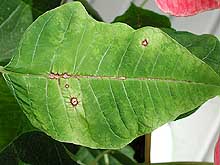
Fig. 13. Scab lesions on a leaf. Note multiple lesions along top of midrib due to channeling of spores in water (click image for larger view).
|
|
The disease affects both leaves and stems of the plant (15). Small, round lesions (1 to 5 mm in diameter) form on the leaf blade, and commonly occur on the midvein or lateral veins where they frequently coalesce (Fig. 13). The spots develop whitish to brown centers, have a dark red to purple rim, and often show a diffuse yellow halo. The most distinctive feature of the spots is that they are buckled out from the upper leaf surface. With time the leaf spots develop a coating of sporulation, causing them to change from white to a velvety brown coloring (Fig. 14). The fungus makes single-celled, ovoid, hyaline conidia (3 to 7 x 1.5 to 4 µm) and also produces larger (7 to 25 x 2.5 to 7 µm), pigmented, 1 to 2 celled conidia, constricted at the septation. The latter spore type, called the “fawcetti conidium” has been thought to be the form most likely to be involved in long-distance spread of the fungus by wind (Fig. 15).
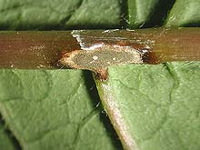
Fig. 14. Sporulating scab lesion on midrib with velvety brown spores (click image for larger view).
|
|
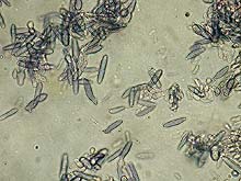
Fig. 15. One to two-celled ‘fawcetti’ conidia of Sphaceloma poinsettiae (click image for larger view). |
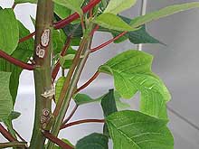
Fig. 16. Scab stem lesions with red pigmentation at margin (click image for larger view).
|
|
The stems of poinsettias show oval to elongated raised cankers, with dimensions of 3 to 10 x 2 to 6 mm. The stem lesions are whitish in color, and there may be red pigmentation around them (Fig. 16). One of the most bizarre features of the disease is the result of a growth regulating chemical produced by the fungus in which a shoot with only a single canker can show “superelongation.” Thus, the internodes are lengthened so that the shoot rises six inches or more above the rest of the crop (Fig. 17). Cassava is affected by a similar fungus, Sphaceloma manihoticola, and that pathogen has been shown to produce gibberellin A4 (62). Even if the lesions on leaves and stems are overlooked, growers will notice the affected shoots that stretch high above the rest of the crop.
|
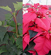
Fig. 17. Superelongation of poinsettia stems in the vegetative stage (left) and later in the flower bract stage (right) due to scab (click image for larger view). |
|
S. poinsettiae is favored by high humidity and wet growing conditions, which are not uncommon during poinsettia propagation. Splashing water will spread the spores easily, and insects might also move spores from plant to plant. Details of the epidemiology of S. poinsettiae have not been determined.
Control studies by Engelhard in Florida two decades ago showed benomyl, chlorothalonil, mancozeb, mancozeb plus thiophanate-methyl and copper hydroxide to be effective against poinsettia scab, whereas dicarboximides were ineffective (25). Trials conducted in 2001 by Chase and Daughtrey (14) indicated that triazoles, strobilurins, materials containing mancozeb, chlorothalonil, and a chlorothalonil plus thiophanate-methyl combination were very effective as protectants against poinsettia scab. Bicarbonates and copper sulfate pentahydrate were less effective but provided some control.
Poinsettia growers are encouraged to scout for scab symptoms on leaves and stems. If scab is detected, infected plants should be removed, and the remainder treated with an effective fungicide. Splashing during irrigation and lengthy periods of leaf wetness should be avoided in order to reduce the opportunity for new scab infections.
|
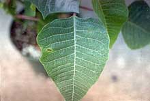
Fig. 18. Alternaria leaf spot on poinsettia (click image for larger view). |
Alternaria leaf spot and blight. Alternaria leaf spot in poinsettia (caused by Alternaria euphorbiicola) was first reported causing commercial losses from Florida in about 1984. In 1986, Yoshimura et al. (59) described what appears to be the same disease caused by Alternaria euphorbiae (=Macrosporium euphorbiae). The disease started in Hawaii in late 1983. Alternaria blight of poinsettias is characterized by small (less than 1 mm in diameter) lesions that are initially water-soaked. The roughly circular lesions turn reddish-brown to dark brown and reach 20 mm in diameter (Fig. 18). Lesions may or may not have a halo. Lesions also form on bracts reaching 40 mm across with a black-purple margin. Stem lesions can result in girdling and subsequent loss of the stem. Cultivar resistance was examined in the 1980s but no work has been done with the multitude of new cultivars introduced since that time. Elimination of water on leaves is important to completely control Alternaria leaf spot of poinsettias. In 1985, iprodione and the combination product of thiophanate methyl and mancozeb were reported effective in controlling Alternaria leaf spot (26). Although both products are still available, McGovern (50) identified azoxystrobin and fludioxonil as giving superior disease control in studies conducted in the late 1990s.
Erwinia blight and cutting rot. Soft rot of poinsettia cuttings was originally described in 1959 from Missouri (55). The pathogen was identified as Erwinia carotovora, the cause of many ornamental soft rot diseases. The onset of symptoms is very rapid with soft rot evident as much as 7 to 10 cm above the cut end of a cutting within 24 hours of infection. The rot starts as a watery area at the cut end or anywhere on the cutting, causing disintegration (Fig. 19). A characteristic rotten fishy odor often is present in the propagation house when this disease is present. Infected cuttings parts that have dried remain sources of active bacteria for at least 6 weeks. Erwinia carotovora is widely distributed throughout most ornamental production areas. It has even been found in irrigation water in some of the southern states. Growers have reported that preventative water treatment with 0.5 to 1 ppm bromine or chlorine has been effective. Use of bactericides is rarely helpful with soft rot diseases of cuttings.
| |
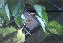
Fig. 19. Erwinia blight on poinsettia cuttings in propagation (click image for larger view).
|
|
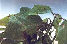
Fig. 20. Angular leaf spots on poinsettia caused by Xanthomonas (click image for larger view). |
|
Xanthomonas leaf spot. Xanthomonas leaf spot on poinsettias was originally reported in 1951 from India (54). It was later described as causing commercial losses in outdoor production of poinsettias in Florida in 1960 (49). The cause is Xanthomonas campestris pv. poinsettiicola. Pinpoint spots start as dull gray to brown slightly water-soaked areas. As the lesions mature they turn yellow to tan and are scattered across the leaf surface. Sometimes they enlarge and become angular in shape due to limited movement across leaf veins (Fig 20). This disease can be confused with early symptoms of scab. Severe infections can cause distortion of new leaves as well as complete chlorosis and finally abscission of older leaves. When the disease was discovered in Florida, researchers found that many, if not all, popular cultivars of the day were highly susceptible to Xanthomonas leaf spot. No new work has been conducted to evaluate cultivar resistance. Control must be based on elimination of all stock plants with Xanthomonas leaf spot. In this day of mass production of rooted poinsettia cuttings it is most important for the propagator to identify a bacterial leaf spot outbreak and eradicate it at the source. Use of copper bactericides may be partially effective in controlling this disease but are rarely effective in stopping an outbreak once infection has occurred. The disease is nearly impossible to control unless plants are produced without overhead watering or exposure to rainfall.
Root Diseases
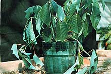
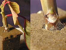
Pythium root rot. Every season, some poinsettia growers encounter crop losses as a result of Pythium root rot. Depending upon the circumstances in the particular greenhouse, a few plants may be affected or a very high percentage of the crop can be lost. Pythium usually attacks early in the season (3), soon after cuttings have been potted. Severely affected rooted cuttings wilt and die rapidly. The base of the cutting is brown and has a water-soaked appearance. The callus and any new roots at the base of the cutting also turn brown. The growth of infected plants that survive is stunted (Fig. 21) and these plants often wilt (Fig. 22) during the heat of the day and recover at night later in the season. Infected roots of established plants are dark brown in color and the outer layers of root tissue strip off leaving a bare strand of inner vascular tissue exposed (Fig. 23). Infected plants that survive until flowering usually flower prematurely and defoliate.
 |
|
 |
|
Fig. 21. Stunting of poinsettia growth (left) due to Pythium root rot compared to healthy plant (right) (click image for larger view). |
|
Fig. 22. Wilted foliage of a poinsettia due to Pythium root rot. Wilted plants may recover at night (click image for larger view).
|
|
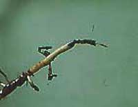
Fig. 23. Roots of poinsettia with dark brown discoloration and sloughed off cortical tissues (click image for larger view).
|
|
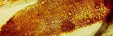
Fig. 24. Spherical oospores of a Pythium sp. in the cortical cells of a poinsettia root (click image for larger view). |
|
An examination of the root cortex cells often reveals the presence of spherical oospores (Fig. 24). However, oospores may be absent and only sparse hyphae are observed in the roots. When such roots are plated onto water agar or a selective medium (21,43), Pythium grows out within 24 hour Pythium irregulare and P. ultimum are common species found in poinsettia roots (29,57) but a recent examination of isolates obtained from clinic samples in Pennsylvania from 1996 to 2001 indicated that Pythium aphanidermatum accounted for 76% of the Pythium root rot cases (Moorman, unpublished). At this time it is not known whether poinsettias are more susceptible to P. aphanidermatum than other species, whether P. aphanidermatum is infecting cuttings during propagation and is being inadvertently shipped to other growers or whether the greenhouse temperatures and other cultural conditions used in poinsettia production make it more likely that P. aphanidermatum will successfully attack poinsettias than other species.
Pythium inoculum may enter the production system from a number of sources. If the propagator of cuttings allows Pythium to infect plants during the callusing and rooting process and infections go unobserved, then Pythium-infected cuttings are sold to customers. Other sources of the organism include contaminated soil from under and between benches or outside the greenhouse that is then moved into pots or irrigation water reservoirs or onto benches or flood floors. If the grower uses contaminated field soil as a component of the potting mix or if the properly treated potting soil is contaminated with untreated soil, it is not unusual for a very high percentage of the crop to be affected. Growers employing potting mixes composed of mixtures of peat moss, bark, vermiculite, coir, peanut or rice hulls, perlite, and other non-soil materials sometimes allow the mix to become contaminated with Pythium-infested soil from the sources noted above. It is known that peat moss can harbor Pythium (45) and it has been found that commercial soilless potting media sometimes contain the pathogen. Surface water supplies such as streams and ponds may bear Pythium. When used as sources of irrigation water, plants will be inoculated regardless of whether the water is applied via sprinkler irrigation, trickle, or by ebb and flow systems. In subirrigation systems, severe cases of waterborne Pythium root rot occur in operations employing long flooding times (more than 30 to 40 minutes) or where flooded floors or benches do not drain completely and pots remain in puddles for extended periods. While it is known that fungus gnats and shoreflies are vectors of Pythium in some systems (31,32,42), the importance of this in potted plant production has not been studied. Factors that favor Pythium root rot development in poinsettias include excessive fertilizer levels (51), high soil moisture levels (2), and soil pHs above 5.5 (1) when P. ultimum is involved. Little work has been done on factors affecting other species known to attack poinsettias.
Pythium root rot is difficult to control once it has begun. Every effort should be directed toward preventing the disease before it begins by eliminating the pathogen from the production system. Pathogen-free potting mixes are essential in this effort and general sanitation practices in the greenhouse should target the removal or treatment of soil that may harbor Pythium. Disinfesting bench surfaces, flood floor systems, potting benches, tools, and equipment that will contact the potting mix is important. If pond or stream water is used for irrigation, the intake pipe should be well above the bottom so that sediment is not drawn in. If the water supply is suspected of being a source of Pythium, it may be necessary to treat the water (22). Irrigation systems in which unused water is recycled, once contaminated with a pathogen, becomes an ongoing source of Pythium. For that reason, ebb and flow system reservoirs should be covered in order to prevent contaminated soil or debris from entering them. To remove plant debris that may harbor the pathogen from the water, the water should be passed over a coarse screen before it is returned to the reservoir.
In a greenhouse operation with a history of Pythium root rot, fungicides or biological control agents should be applied as early in the cropping cycle as possible. Biological agents including species and strains of Trichoderma, Bacillus, Gliocladium, and Streptomyces, can be applied to the potting mix before, during or immediately after transplant. In general it is recommended that chemical pesticides not be applied to the potting mix during the period from 10 days before to 10 days after applying the biological control agent. Biological control agents and fungicides may have to be applied more than once in order to maintain adequate protection throughout the season, particularly on stock plants. Several fungicides, including mefenoxam/metalaxyl, propamocarb, and etridiazole are registered for use on poinsettias in the U.S. However, some populations of P. aphanidermatum and P. irregulare have resistance to mefenoxam/metalaxyl.
Phytophthora root, crown, leaf, bract, and flower blight. Two species of Phytophthora, P. nicotianae Breda de Haan (=P. parasitica Dastur) and P. drechsleri have been recovered from poinsettias in greenhouses at various locations in the United States. Root, crown, and stem rot caused by P. nicotianae was first described by Engelhard and Ploetz in 1979 (27), and foliar infection caused by both P. nicotianae and P. drechsleri was first described by Yoshimura et al. in 1985 (58). Infected roots are brown and depending on the environmental conditions and the age of the plant, infection may be present for varying lengths of time before wilt or stunting is noticed. Infections at and above the soil line are characterized by purple-black lesions that may expand rapidly from stems to brack petioles causing the bracts to wilt (Fig. 25). Leaf lesions are paper-like and dry in texture, grayish brown at first, turning brown to black. Blight symptoms produced by these two species are virtually identical (58).
|
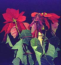
Fig. 25. Wilting of flower bracts and foliage due to stem lesions caused by a Phytophthora sp. (click image for larger view).
|
|
Both P. nicotianae and P. drechsleri are heterothallic, requiring A1 and A2 mating types to complete the sexual stage, and form thick-walled oospores (28). Single isolates readily produce sporangia on diseased tissue and sporangia can quickly release swimming zoospores as the crop is watered. Recent epidemics of Phytophthora blight in floriculture production facilities throughout the U.S. were caused by the spread of a single clonal lineage (e.g., sporangia and zoospores from a single Phytophthora isolate) including one epidemic in poinsettia caused by P. drechsleri (Lamour and Hausbeck, unpublished). Many poinsettia production facilities utilize ebb and flood watering strategies which provide frequent opportunities for the spread of sporangia and zoospores to other plants as the crop is watered. The incubation period between the initial infection of a plant and subsequent visible symptoms provides ample time for spread of inoculum throughout a facility.
Control strategies that prevent initial infections are most effective in limiting the development of Phytophthora epidemics in poinsettia. These include the use of pathogen-free potting mix and strict sanitation in the production facility. The fungicides metalaxyl (Subdue) and more recently mefenoxam (Subdue Maxx) have been shown to be very effective in controlling Phytophthora blight of poinsettia (58). However, isolates of P. nicotianae resistant to the highest rate of application of metalaxyl or mefenoxam have been reported from floriculture production facilities (30, Lamour and Hausbeck, unpublished). Applying fungicide to known infected plants will increase the chance of selecting for insensitive isolates. The most effective way to halt an epidemic is to remove all plants from a production area in a facility that have been irrigated with water from the reservoir that collects recycled water from known infected plants. Even healthy looking plants may be infected so strict sanitation following an epidemic is mandatory to prevent re-occurrences.
Rhizoctonia stem rot. Stem rot caused by Rhizoctonia solani Kuhn AG-4 is the most important disease that affects poinsettia during propagation. The pathogen has a very wide host range, as well as a high competitive saprophytic ability. This combination of characteristics provides an ideal opportunity for both infection of host tissue and colonization of plant debris followed by subsequent long-term survival either as mycelium or sclerotia associated with crop debris. Growers may practice strict sanitation but often times R. solani becomes an indigenous pathogen in greenhouse production facilities as the fungus survives between crops in infested debris left behind on the bench, equipment, floor, walkways, and even in crevasses on wooden benches.
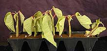
Poinsettia may be propagated in 10-unit polyfoam rooting strips, rockwool cubes, or stuck directly in a soilless potting mix in pots. Propagation of poinsettia typically occurs in the hot months of July and August in the greenhouse under conditions of almost constant leaf wetness from mist systems designed to prevent the cutting from wilting. Under these ideal conditions, Rhizoctonia stem rot develops when debris colonized by R. solani is dispersed to the rooting strip by splashing water or other means of contact. The initial symptoms of disease are small lesions that often develop at the point on the stem even with the top surface of the rooting cube. Lesions have a dry appearance with a dark border and tan center (Fig. 26). Multiple lesions may develop on a single cutting. If newly made cuttings become infected, lesions expand rapidly girdling the stem and collapsing the cutting within 5 to 7 days (Fig. 27). Cuttings that have been in propagation a week or more are more resistant to R. solani. Drooping leaves on cuttings that contact the bench surface also can be infected by Rhizoctonia harbored in residual debris. Infected areas on leaves are brown and beads of white latex from the host often appear on the infected tissue under conditions of high humidity. Infected leaves are quickly colonized and provide an avenue of entry to stems for further stem rot development. Commercial ELISA kits are available for growers and diagnosticians to detect R. solani in infected poinsettia tissue (5). Once one cutting in a rooting strip becomes infected, R. solani can use that diseased tissue as a food base to grow both on the surface and through the strip to infect other cuttings in the 10-unit strip. Sclerotia and hyphae may be visible on the surface of the rooting cube particularly on the sides exposed when the protective cover holding the strip is removed (Fig. 28).
 |
|
 |
|
Fig. 26. Poinsettia cutting with stem lesion in leaf abscission area (left) and close up of Rhizoctonia hyphae growing out into rooting cube (right) (click image for larger view). |
|
Fig. 27. Collapse of a poinsettia cutting with stem rot in a rooting strip. Other cuttings in strip have visible stem lesions as well (click image for larger view). |
|
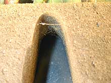
Fig. 28. Rooting strip removed from protective plastic cover to reveal Rhizoctonia hyphae growing through strip (click image for larger view).
|
|
Rhizoctonia crown and root rot. Root rot may develop either in the rooting cube or on rooted cuttings transplanted to pots as the crop is finished for retail. Infected roots become water-soaked then brown. Both root tips and sections of the root away from the tip may develop symptoms. Crown rot can develop on the stem as lesions expand from stem infections occurring during propagation. However, stem lesions may develop at a much slower pace on rooted plants, since this tissue is more hardened off and thus more resistant than stems of newly made cuttings. Foliar symptoms of crown and root rot include chlorosis, leaf necrosis, wilting, defoliation, and plant death, but often the most common symptom is stunting. Root rot infections may be initiated from lesions on stems or from inoculum introduced to the potting mix from debris surviving in the greenhouse. Generally, moist but not wet conditions in the potting mix favor development of Rhizoctonia crown and root rot on potted plants. Spacing plants with a full canopy too close together can result in moisture and soil temperatures favorable for development of disease due to shading of the container surface.
Control of stem and root rot begins with thorough removal of all crop debris from the production facility at the end of a cropping cycle. Sanitation of work area and bench surfaces with surface disinfectants is important. During propagation, misting cycles should be monitored closely to avoid over wetting foliage of cuttings once newly made cuttings become turgid. Daily scouting of propagation areas is needed so that rooting strips with any cutting showing symptoms or signs of stem rot can be removed. No rooted cuttings from a strip with an infected cutting should be transplanted since the pathogen may have grown through the foam cubes resulting in apparently healthy but infected cuttings that will not produce saleable plants.
In greenhouse production facilities with a history of Rhizoctonia stem and root rot, soaking dry rooting strips in a fungicide solution can protect cuttings from disease (4). Fungicides such as flutolanil, iprodione, and chlorothalonil actually prevented R. solani from colonizing the rooting cube (4). Fludioxonil and the new strobilurins such as azoxystrobin also are effective for stem rot control (7). Generally one application of fungicide is sufficient to protect the crop during the propagation cycle. Since some fungicides affect the number of roots formed on cuttings, growers should test specific fungicide by cultivar combinations for safety before using a product on the entire crop (6). After transplanting, fungicide drenches may be needed at regular intervals to prevent crown and root rot.
In addition to traditional fungicides, several biocontrol agents such as Burkholderia cepacia, Paecilomyces lilacinus, and binucleate Rhizoctonia spp. (BNR) have shown potential for control of Rhizoctonia stem and root rot (11). Commercial biocontrol agents mentioned above also are available, although those based on Gliocladium virens are phytotoxic to unrooted poinsettia cuttings (Benson, unpublished). In a unique study, a sequential application of B. cepacia to rooting strips followed by incorporation of a Pesta formulation of BNR into potting mix at transplanting protected poinsettia from stem rot in propagation and root and stem rot for 52 days after transplanting (38).
Conclusion
The long production season for poinsettias (from propagation in the hot months of summer to vegetative growth and then flower bract development in the shorter days and cooler months of fall and early winter) provides a wide range of environmental conditions that can foster a series of diseases. We have described the major poinsettia diseases that are widespread in the industry. A number of other less common diseases can cause significant problems for individual growers when favorable environmental conditions prevail. In addition to biotic agents, improper fertilization practices can cause symptoms in poinsettias. A convenient table summarizing the management of poinsettia diseases and nutrient deficiencies is available online from Penn State University. A description including images of physiological disorders in poinsettia due to improper fertilization can be found online from North Carolina State University. Plant pathologists will continue research and extension efforts to provide growers with the best possible disease management strategies that will help to preserve the poinsettia as the Christmas flower.
Electronic Resources for Further Information
Diseases of poinsettia: Common Names of Plant Diseases
Diseases and Disorders of Poinsettia
Plant Disease Facts: Poinsettia Diseases
Paul Ecke Ranch, Technical Information Bulletin: Poinsettia Scab
Poinsettia Scab (Spot Anthracnose)
Texas Poinsettia Producers Guide
Literature Cited
1. Bateman, D. F. 1962. Relation of soil pH to development of poinsettia root rots. Phytopathology 52:559-566.
2. Bateman, D. F. 1963. Factorial analysis of environment and pathogens in relation to development of the poinsettia root rot complex. Phytopathology 53:509-516.
3. Bateman, D. F., and Dimock, A. W. 1959. The influence of temperature on root rots of poinsettia caused by Thielaviopsis basicola, Rhizoctonia solani, and Pythium ultimum. Phytopathology 49:641-647.
4. Benson, D. M. 1991. Control of Rhizoctonia stem rot of poinsettia during propagation with fungicides that prevent colonization of rooting cubes by Rhizoctonia solani. Plant Dis. 75:394-398.
5. Benson, D. M. 1992. Detection by enzyme-linked immunosorbent assay of Rhizoctonia species on poinsettia stem cuttings. Plant Dis. 76:578-581.
6. Benson, D. M. 1992. Fungicides as foliar sprays or rooting cube soaks in propagation of poinsettia. HortScience 27:1006-1008.
7. Benson, D. M., and Parker, K. C. 2001. Efficacy of Heritage 50W for control of Rhizoctonia stem rot of poinsettia, 2000. APS Press, Fungic Nematic Tests OT238. Byrne, J. M., and Hausbeck, M. K. 1998. Influence of temperature and relative humidity on infection processes and sporulaton of Oidium sp. on poinsettia foliage. (Abstr.) Phytopathology 88:S134.
9. Byrne, J. M., and Hausbeck, M. K. 1999. The effect of temperature on sporulaton of Oidium sp. on poinsettia foliage. (Abstr.) Phytopathology 89:S10.
10. Byrne, J. M., Hausbeck, M. K., and Shaw, B. D. 2000. Factors affecting concentrations of airborne conidia of Oidium sp. among poinsettias in a greenhouse. Plant Dis. 84:1089-1095
11. Cartwright, D. K., and Benson, D. M. 1995. Biological control of Rhizoctonia stem rot of poinsettia in polyfoam rooting cubes with Pseudomonas cepacia and Paecilomyces lilacinus. Biol. Contr. 5:237-244.
12. Celio, G. J., and Hausbeck, M. K. 1997. Evaluation of poinsettia cultivars for susceptibility to powdery mildew. HortScience 32:259-261.
13. Celio, G. J., and Hausbeck, M. K. 1998. Conidial germination, infection structure formation, and early colony development of powdery mildw on poinsettia. Phytopathology 88:105-113.
14. Chase, A. R., and Daughtrey, M. L. 2001. Control of poinsettia scab in 2001. Pages 3-5 in: Proceedings, 17th Annual Conference on Insect and Disease Management on Ornamental Plants, Society of American Florists, Alexandria, VA.
15. Daughtrey, M. 2001. Disease Doctor: What are those bumpy spots on my poinsettia leaves? Greenhouse Business 7(6):35-36.
16. Daughtrey, M. and Hall, J. 1992. Powdery mildew-a new threat to your poinsettia crop. Grower Talks, Sept:23-31.
17. Daughtrey, M., and Macksel, M. T. 1994. Efficacy of fungicides for control of powdery mildew disease of poinsettia. 1992. Fungic Nematic. Tests 49:359.
18. Daughtrey, M. and Tobiasz, M. 2000. Evaluation of fungicides for control of powdery mildew of poinsettia. Fungic. Nematic. Tests 55:559.
19. Daughtrey, M. and Tobiasz, M. 2001. Evaluation of fungicide programs for control of powdery mildew of poinsettia, 2000. Fungic. Nematic. Tests 2001:0T25.
20. Ecke, P. Jr., Matkin, O. A., and Hartley, D. E. 1990. The Poinsettia Manual. Paul Ecke Poinsettias. California.
21. Eckert, J. W., and Tsao, P. H. 1962. A selective antibiotic medium for isolation of Phytophthora and Pythium from plant roots. Phytopathology 52:771-777.
22. Ehret, D. L., Alsanius, B., Wohanka, W., Menzies, J. G., and Utkhede, R. 2001. Disinfestation of recirculating nutrient solutions in greenhouse horticulture. Agronomie 21:323-339.
23. Elad, Y., Yunis, H., and Katan, T. 1992. Multiple resistance to benzimidazoles, dicarboximides, and diethofencarb in field isolates of Botrytis cinerea in Israel. Plant Pathol. 41:41-46.
24. EmilyCompost.com. 2001. Joel Roberts Poinsett (1779 - 1851). Online. Holiday interests section.
25. Engelhard, A. W. 1983. Control of poinsettia scab with fungicides. Fungic. Nematic. Tests 38:182.
26. Engelhard, A. W. 1985. Alternaria leaf spot and blight of poinsettia. Florida Nurs. 32(12):40-41, 44.
27. Engelhard, A. W., and Ploetz, R. C. 1979. Phytophthora crown and stem rot, an important new disease of poinsettia (Euphorbia pulcherrima). Proc. Fla. State Hort. Soc. 92:348-350.
28. Erwin, D. C., and Ribeiro, O. K. 1996. Phytophthora Diseases Worldwide. American Phytopathological Society, St. Paul, MN.
29. Farr, D. F., Bills, G. F., Chamuris, G. P., and Rossman, A. Y. 1989. Fungi on plants and plant products in the United States. St. Paul: APS Press.
30. Ferrin, D. M., and Kabashima, J. N. 1991. In vitro insensitivity to metalaxyl of isolates of Phytophthora citricola and P. parasitica from ornamental hosts in southern California. Plant Dis. 75:1041-1044.
31. Gardiner, R. B., Jarvis, W. R., and Shipp, J. L. 1990. Ingestion of Pythium spp. by larvae of the fungus gnat Bradysia impatiens (Diptera, Sciaridae). Ann. Appl. Biol. 116:205-212.
32. Goldberg, N. P., and Stanghellini, M. E. 1990. Ingestion-egestion and aerial transmission of Pythium aphanidermatum by shore flies (Ephydrinae: Scatella stagnalis). Phytopathology 80:1244-1246.
33. Hausbeck, M. K., and Pennypacker, S. P. 1991. Influence of grower activity and disease incidence on concentrations of airborne conidia of Botrytis cinerea among geranium stock plants. Plant Dis. 75:798-803.
34. Hausbeck, M. K., and Pennypacker, S. P. 1991. Influence of grower activity on concentrations of airborne conidia of Botrytis cinerea among geranium cuttings. Plant Dis. 75:1236-1243.
35. Hausbeck, M., Kalishek, J., Daughtrey, M., and Barnes, L. 1994. Keep the colonies at bay, part 1. Greenhouse Grower 12(9):45, 47-48.
36. Hausbeck, M., Kalishek, J., Daughtrey, M., and Barnes, L. 1994. Keep the colonies at bay, part 2. Greenhouse Grower 12(10):88, 91-92.
37. Hausbeck, M. K., and Moorman, G. W. 1996. Managing Botrytis in greenhouse-grown flower crops. Plant Dis. 80:1212-1219.
38. Hwang, J. and Benson, D. M. 2001. Biocontrol of Rhizoctonia stem and root rot of poinsettia with integrated application of Burkholderia cepacia and Binucleate Rhizoctonia. Plant Dis. 85 (in press).
39. Jarvis, W. R. 1960. An apparatus for studying hygroscopic responses in fungal conidiospores. Trans. Br. Mycol. Soc 43:525-528.
40. Jarvis, W. R. 1962. Splash dispersal of spores of Botrytis cinerea Pers. Nature (London) 193:599.
41. Jarvis, W. R. 1977. Botryotinia and Botrytis species: taxonomy, physiology, and pathogenicity. Vol. 15, Canada Department of Agriculture Monographs. Ottawa: Canada Department of Agriculture.
42. Jarvis, W. R., Shipp, J. L., and Gardiner, R. B. 1993. Transmission of Pythium aphanidermatum to greenhouse cucumber by the fungus gnat Bradysia impatiens (Diptera, Sciaridae). Ann. Appl. Biol. 122:23-29.
43. Jeffers, S. N., and Martin, S. B. 1986. Comparison of two media selective for Phytophthora and Pythium species. Plant Dis. 70:1038-1043.
44. Jenkins, A. E. and Ruehle, G. D. 1942. A new species of Sphaceloma-On poinsettia. Proc. Biol. Soc. Wash. 55:83-84.
45. Kim, S. H., Forer, L. B., and Longenecker, J. L. 1975. Recovery of plant pathogens from commercial peat products. Proceedings of the. American Phytopathological Society 2:124.
46. LaMondia, J. A., and Douglas, S. M. 1997. Sensitivity of Botrytis cinerea from Connecticut greenhouses to benzimidazole and dicarboximide fungicides. Plant Dis. 81:729-732.
47. Lee, I.-M., Klopmeyer, M., Bartoszyk, I. M., Gundersen-Rindal, D. E., Chou, T.-S., Thomson, K. L. and Eisenreich, R. 1997. Phytoplasma induced free-branching in commercial poinsettia cultivars. Nature Biotechnology 15:178-182.
48. Lee, I.-M. 2000. Phytoplasma casts a magic spell that turns the fair poinsettia into a Christmas showpiece. Online. Plant Health Progress doi:10.1094/PHP-2000-0914-01-RV.
49. McFadden, L. A., and Mowry, H. R. 1962. Bacterial leaf spot disease of poinsettia in Florida. Plant Dis. Rep. 46:551-554.
50. McGovern, R. J. 1998. Evaluation of fungicides for control of Alternaria leaf spot and blight and Choanephora wet rot in poinsettia, 1998. Fungic.and Nematic. Tests 54:547.
51. Moorman, G. W. 1986. Increased mortality caused by Pythium root rot of poinsettias associated with high fertilization rates. Plant Dis. 70:160-162.
52. Moorman, G. W., and Lease, R. J. 1992. Benzimidazole- and dicarboximide-resistant Botrytis cinerea from Pennsylvania greenhouses. Plant Dis. 76:477-480.
53. Northover, J., and Matteoni, J. A. 1986. Resistance of Botrytis cinerea to benomyl and iprodione in vineyards and greenhouses after exposure to the fungicides alone or mixed with captan. Plant Dis. 70:398-402.
54. Patel, M. K., Bhatt, V. V., and Kulkarni, Y. S. 1951. Three new bacterial diseases of plants from Bombay. Indian Phytopathol. 4:144.151.
55. Rogers, M. A. 1959. Decay of poinsettia cuttings by the soft rot bacterium, Erwinia carotovora (Jones) Holland. Plant Dis. Rep. 43:1236-1238.
56. Ruehle, G. D. 1941. Poinsettia scab caused by Sphaceloma. Phytopathology 31:947-948.
57. Tompkins, C. M., and Middleton, J. T. 1950. Etiology and control of poinsettia root and stem rot caused by Pythium spp. and Rhizoctonia solani. Hilgardia 20:171-182.
58. Yoshimura, M. A., Uchida, J. Y., and Aragaki, M. 1985. Etiology and control of Poinsettia blight caused by Phytophthora nicotianae var. parasitica and P. drechsleri. Plant Dis. 69:511-513.
59. Yoshimura, M. A., Uchida, J. Y., and Aragaki, M. 1986. Etiology and control of Alternaria blight of poinsettia. Plant Dis. 70:73-75.
60. Yourman, L. F., Jeffers, S. N., and Dean, R. A. 2001. Phenotype instability in Botrytis cinerea in the absence of benzimidazole and dicarboximide fungicides. Phytopathology 91:307-315.
61. U. S. Department of Agriculture, 2001. Floriculture Crops 2000 Summary, National Agricultural Statistics Service, Agricultural Statistics Board.
62. Ziegler, R. S. 1982. The Superelongation Disease of Cassava: Pathogen Taxonomy, Gibberellin Production, and Some Characteristics of Host Resistance, PhD Thesis, Cornell University.

