Melissa B. Riley1, Margaret R. Williamson1, and Otis Maloy2
1Department of Plant Pathology and Physiology, Clemson University, Clemson, SC
2Department of Plant Pathology, Washington State University, Pullman, WA
Riley, M.B., M.R. Williamson, and O. Maloy. 2002. Plant disease diagnosis. The Plant Health Instructor. DOI: 10.1094/PHI-I-2002-1021-01
Spanish Version
A plant pathologist or a student taking plant pathology is often asked by friends or associates the following questions. What is wrong with my plant; followed by, what can I do to get rid of the problem? It may be too late to help the specific plant when the question is asked, but proper diagnosis may be extremely important in preventing the problem on other plants or in preventing the problem in the future.
How does a plant pathologist or a plant pathology student go about diagnosing plant problems? The diagnostician must have very good observation skills, and s/he also needs to be a good detective. It is important to keep an open mind until all of the facts related to the problem can be collected. The possibility of multiple causal factors must also be considered.
Control measures depend on proper identification of diseases and of the causal agents. Therefore, diagnosis is one of the most important aspects of a plant pathologist's training. Without proper identification of the disease and the disease-causing agent, disease control measures can be a waste of time and money and can lead to further plant losses. Proper disease diagnosis is therefore vital.
Often, plant pathologists have to rely on symptoms for the identification of a disease problem. For example, Dr. Shoe is asked by Ms. Green to examine azaleas in her nursery. When Dr. Shoe gets to the nursery, he observes that the azaleas in Greenhouse 1 are wilted. When he removes plants from their pots, the roots appear to be rotten. Ms. Green wants to know right away what she should do with the azaleas in Greenhouse 2 where no wilting is currently being observed. Dr. Shoe is asked to make recommendations even before he has looked at plants in the second greenhouse. Because similar symptoms can be produced in response to different causal agents, the use of symptoms alone is often an inadequate method for disease identification. The identification of the disease-causing agent may take a week or more. What can Dr. Shoe do for Ms. Green now?
One of the most important things is for Dr. Shoe to use his powers of observation. He needs to ask many questions related to the azalea's care and culture in order to eliminate or identify possible causes of the problem. He also needs to consider various environmental and cultural factors. As a result of his questions and observations he may:
- Be able to identify a disease and disease-causing agent,
- Be able to narrow the problem down to several possibilities which will require further study in the laboratory before he can make a final diagnosis, or
- Be completely baffled by the problem.
Regardless of the outcome, Ms. Green still expects a recommendation as to what she should do now.
This article presents the various steps/activities which are associated with accurate plant disease diagnosis. The process may vary with different diseases and conditions but the overall process is relatively consistent. The steps all require careful observations and questions. The steps include:
Know What is Normal
Proper plant identification. Identification of affected plants is one of the first steps in diagnosing a plant disease. Both scientific and common names of the plant should be noted. Common names should not be relied upon since some distinctly different plant species may have the same common name, and the common name used in one area may be used for a completely different species in another area. The common name "vinca" has been used to describe plants belonging to two different genera, Vinca, a perennial, and Catharanthus, an annual. Another example is "monkey grass" which is used to describe Liriope and Ophiopogon (mondo grass). An example from forestry is "cedar" which is used to describe eastern red cedar (Juniperus), western red cedar (Thuja), Port Orford cedar (Chamaecyparis), incense cedar (Libocedrus), and Atlas cedar (Cedrus). Obviously the use of common names can cause confusion in identification and recognition of problems.
In addition to knowing the common and scientific names of an affected plant, it is important to know the specific variety or cultivar, whenever possible. A great variation in susceptibility to a specific disease may occur within different cultivars of a plant species. For example, when we look at the susceptibility of wheat to wheat stem rust caused by Puccinia graminis f. sp. tritici, we know that all wheat cultivars are not susceptible to all races of P. graminis. The major control measure for this disease is based on planting wheat cultivars each year that are resistant to the pathogen races that are predicted to be present during the growing season. Tomato cultivars having the "Better Boy" genetic background are generally resistant to root-knot nematodes while those with the genetic background of the variety "Rutgers" are susceptible, so knowing the genetic background of a cultivar can be important. Knowing the cultivar and its susceptibility to various diseases can narrow down the possible disease agents to consider.
Knowing the identity of the plant species affected allows the pathologist to utilize various resources that contain lists of plant diseases associated with specific plants. These lists are very helpful in suggesting possible pathogenic agents. An example of such a list is found on the American Phytopathological Society (APS) web site as a part of its the online resources. Once you select the plant of interest, you will see a list of bacterial, fungal, nematode, parasitic plants and viral diseases associated with the specific plant. Westcott's Plant Disease Handbook is useful because specific symptoms are associated with each disease7. APS Press has published a list of fungal diseases and hosts4. This book followed a previous publication by USDA12, but the APS Press publication only includes fungal pathogens. The original USDA publication, while now somewhat outdated, included fungal, bacterial, viral and nematode pathogens, as well as physiological problems. Other resources are available which include the APS Press Compendium series on diseases and disorders for specific plant species, such as roses8 or diseases for specific regions, such as Florida, USA2. In some cases these lists of plant diseases may suggest potential disease possibilities or they may lead the diagnostician to rule out other diseases. One factor to keep in mind, however, is that these lists are often incomplete or the disease may be new and unreported on the plant or in a specific region. The best possible option is to utilize several different resources since one may not have a complete index of potential diseases on a specific plant.
Recognize healthy plant appearance. It is important to know the normal appearance of the plant species you are investigating. Each plant species has special growth habits, colors and growth rates. If you do not know what to expect of the plant you cannot recognize when something is wrong. Does the plant normally have new foliage that is yellow or red and becomes darker green as the foliage ages? Many ornamental shrubs have been developed and marketed for the ornamental value of such brightly colored new growth. These plants are highly prized for this coloration; however, if an individual does not know that this coloration is the normal appearance of the plant, s/he may think that the plant is diseased. It is important to know what the normal appearance of a plant is before you decide there is a problem. It is also important to remember that appearance can vary with different cultivars. Some plant cultivars have naturally yellow to pale green leaves (e. g. new hosta cultivars, herbs like golden oregano, and coleus varieties) which at first glance appear to have symptoms of under-fertilization, root stress or soil pH problems.
Once the "normal" appearance of the specific plant is determined, several comparisons can be made between the problem plants and healthy plants. Compare characteristics such as overall size, shape, and coloration; leaf shape, size, coloration, and distribution; root distribution and coloration; and bark, stem or trunk texture and coloration. It is also important to note normal events, such as leaf drop, that may occur in a healthy plant. For example, some holly species normally drop leaves in the spring.
The affected parts of the plants should also be noted. Are there symptoms on the roots, leaves, stems, flowers, or fruit? Is the entire plant involved? Is only one limb or side of a plant involved? Answers to these questions can assist in the identification of the problem.
Check for Symptoms and Signs
Identify characteristic symptoms. Describing the characteristic symptoms exhibited by a specimen can be very difficult to do accurately. Because of this, it is often difficult, if not impossible, to determine what is wrong with a plant when a person is describing symptoms over the phone. As a test of this you may want to take a plant exhibiting symptoms and have three different individuals describe the symptoms that they observe on a sheet of paper. Next, compare the descriptions. Do the descriptions vary significantly? Could you visualize the symptoms by the way any one of the individuals described the diseased plant? Symptoms can often be grouped as follows; for definitions of terms, see the APSnet Education Center Illustrated Glossary:
- Underdevelopment of tissues or organs. Examples include such symptoms as stunting of plants, shortened internodes, inadequate development of roots, malformation of leaves, inadequate production of chlorophyll and other pigments, and failure of fruits and flowers to develop.
- Overdevelopment of tissues or organs. Examples include: galls on roots, stems, or leaves, witches' brooms, and profuse flowering.
- Necrosis or death of plant parts. These may be some of the most noticeable symptoms, especially when they affect the entire plant, such as wilts or diebacks. Other examples include shoot or leaf blights, leaf spots, and fruit rots.
- Alteration of normal appearance. Examples include mosaic patterns of light and dark green on leaves, and altered coloration in leaves and flowers.
Diseases also involve a progression of symptoms that can vary significantly. The progression of symptoms is one of the most important characteristics associated with problems caused by biotic agents. Diseases can result in primary and secondary symptoms. For example, decayed roots on a tree may be a primary symptom while the toppling over of the tree or windthrow is a secondary symptom. At later stages of a disease, secondary invaders may also obscure the original disease symptoms so that symptoms observed at the later stages of the disease are not typical of the symptoms developed in response to the original pathogen.
It is important to look for a progression of disease symptoms in plants exhibiting problems. In some cases, such as improper herbicide usage, symptoms observed may be similar to spots present as a result of an infectious agent. The difference is that with herbicide injury, the symptoms usually appear suddenly and there is no observable progression of symptoms. The spots may also follow spray patterns of the herbicide. Herbicides, such as 2,4-D, can cause leaf distortion which may be confused with viral diseases. However, when new leaves form, they will generally be free of symptoms, indicating a lack of symptom progression.
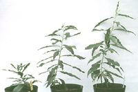
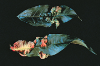
Identify symptom variability. Variations in symptoms expressed by diseased plants may lead to an improper diagnosis. These variations can result from a couple of factors. It is possible that there is more than one problem present, and in some cases there may be more than one pathogen infecting a plant. Symptoms associated with these infected plants may be significantly different from the symptoms expressed in response to each of the different pathogens acting separately. The disease symptoms exhibited by multiple pathogens infecting a plant may be either more severe or less severe than if the plant were infected with just one of the pathogens. This is commonly seen in multiple infections due to viruses. An example of this is shown in Figure 1 which shows peach seedlings infected with single or multiple viruses. The seedling on the left is infected with both Prune dwarf virus and Prunus necrotic ringspot virus. The seedling in the middle is infected with Prune dwarf virus alone and the seedling on the right is infected with Prunus necrotic ringspot virus alone.
|
| Figure 1. Peach seedlings infected with various viruses alone or in combination. Peach seedlings infected with both Prune dwarf virus and Prunus necrotic ringspot virus (seedling on left), infected with Prune dwarf virus (seedling in middle) and infected with Prunus necrotic ringspot virus (seedling on right). (Used by permission of S. Scott) |
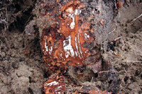
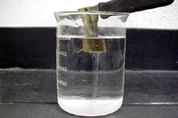
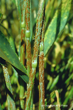
Look for signs of biotic causal agents. Signs of plant disease agents are the observable evidence of the actual disease-causing agent. Signs may include the mycelia of a fungal agent, fungal spores, and spore-producing bodies. Indications of insects causing problems may include the actual insect, insect frass, mite webbing, and insect eggs. Signs are much more specific to disease-causing agents than are symptoms and are extremely useful in the diagnosis of a disease and identification of the agent causing the disease. The use of a hand lens and a knife can be valuable for a diagnostician in the field. Cutting into the bark of ornamental plants and trees at the soil surface may lead to the observation of mycelial mats of root rot fungi such as Armillaria spp. (Figure 2). Bacterial ooze can be observed by cutting stems and placing them in water (Figure 3). Masses of different spores such as rust spores (Figure 4) on leaves can also be important in disease diagnosis. Powdery mildews are typically diagnosed by the observation of the gray to white mycelia and conidia observed on the surface of leaves and flowers (Figure 5).
|
|
 |
|
| Figure 2. White mycelia of Armillaria growing under bark of peach tree. (Used by permission of G. Schnabel) |
|
Figure 3. Bacterial ooze from cut tomato stem infected with Ralstonia solanacearum. (Used by permission of M. Williamson) |
|
|
|
| |
|
|
 |
|
 |
|
| Figure 4. Stem rust on barley. caused by Puccinia graminis. (Courtesy B.Steffenson) |
|
Figure 5. Powdery mildew on apple blossom cluster caused by Podospaera leucotricha. (Courtesy K D. Hickey) |
|
Dissecting and compound microscopes are useful for the observation of specific spores and spore structures, and can lead to further identification of possible disease agents. Knowledge concerning the use of microscopes and a hand lens is vitally important to the diagnostician. Signs of plant disease agents can often be overlooked unless careful observations are conducted. Signs are not visible when taking a quick ride by plants looking through the windshield of a truck and may not even be visible to the naked eye.
Identify Plant Part Affected - Are symptoms associated with specific plant parts?
It is important to note if the symptoms observed are associated with specific plant parts. For example, is a wilt observed correlated with a disruption of the vascular system which may be indicated by browning of the vascular system or are the roots of the plants abnormal including rots, decreased feeder roots, etc.; are necrotic lesions observed strictly on younger leaves? The symptoms of some diseases are most commonly seen on specific plant parts and this observation can be important in diagnosis.
Observe Patterns
Check distribution of symptoms. One of the first things that a diagnostician should note is how the diseased plants are distributed over the affected area. Are they distributed uniformly across an area or are they localized? Is there a definite pattern to the distribution? For example, does it occur only along the edges of a greenhouse near open windows, next to roadways or driveways, in low spots of a field, along a planted row, or is it affecting plants at random in a field? This distribution can be especially important in looking at the possibility of non-infectious problems, such as improper herbicide use or various soil factors11. A uniform pattern on an individual plant and uniform damage patterns over a large area are generally not associated with biotic agents, but are usually due to abiotic agents.
How prevalent is the problem? Are all plants affected? Infectious problems generally occur over time and there is a progression of symptoms. Rarely will all of the plants be affected. Generally, disease problems caused by biotic agents will be observed when they are causing problems on a low percentage of plants at least at the start of the disease, unless there were extenuating circumstances, such as the use of infected seeds. Even then, rarely will 100% infection be observed. When a problem appears in 100% of the plants, it more commonly results from factors such as soil conditions (deficiencies or toxicities), adverse climatic factors (cold temperatures, hail, drought, etc.), or toxic chemicals (improper pesticide use, growth regulators, air pollutants, such as ozone, etc.).
What has been the progression of symptoms on plants in the affected area? If the symptoms all appeared at the same time and there has been no further development of symptoms, this would indicate a possible episodic event such as a change in temperature or possible improper chemical usage. If however, the symptoms started in one area and slowly spread to other areas and the severity of disease symptoms changed over time, this would be more indicative of the presence of a biotic agent. Biotic agents can also include insects and mammals, such as voles, which may be feeding on plants in an area.
Check for host specificity. Is the problem occurring in only one plant species or are different plant species affected? If different plant species are affected, this suggests the possibility of a non-infectious problem which could be related to cultural or environmental problems. However, Phytophthora and Pythium root rots can cause problems on many different plant species; therefore, the fact that more than one plant species is affected does not completely eliminate infectious agents. If there is more than one species of plant involved, are these plants closely related and can they be infected by a common pathogen?
Ask Questions
Review the cultural practices and growing environment. It is vital that a diagnostician question the activities that have been conducted around the affected plants. The problem may not be due to anything that the grower has done; the problem could be related to what his/her neighbor has done. Information pertaining to the growing environment to which the affected plant has been exposed is a vital piece of the puzzle. It is especially important to document changes in the environment. Environmental factors to consider include: extreme temperatures (freezing and heat), rainfall, hail, lightning, prolonged drought, temperature inversions (important in possible air pollutant damage and pesticide drift) and prevailing winds. All of these abiotic factors can be important to the problem. Site factors such as soil type, possible drainage problems, and soil pH should also be evaluated.
Cultural and maintenance activities can be significant. What pesticides or other chemicals have been applied? At what rate and when were they applied? Who applied the chemicals? What equipment was used in its application? What other activities have occurred? Has someone been mowing in the area? Has the highway department been working along the roadway, possibly applying herbicides? Have any unusual occurrences or weather patterns been noted? Many times careful investigation by the diagnostician is required because, in some cases, someone may have done something improperly and may be unwilling to admit their error.
Laboratory Tests
Sometimes neither symptoms nor signs provide enough specific or characteristic information to decide the cause of an infectious plant disease. In such cases, it may be necessary to bring a sample back to the laboratory for further tests to isolate and identify the causal agent. This can be a time-consuming and labor-intensive process that takes specialized skills.
Incubation of plant material. One of the first steps when getting back to the laboratory may be to place a sample of the diseased tissue under conditions that will allow an infectious agent to grow and possibly induce sporulation. This can be accomplished by placing a leaf in a moist chamber11,13. A moist chamber can be a sterile petri dish containing a wet filter paper in the bottom of the dish and a triangle of glass tubing on which the sample is placed so that the sample is not directly on the wet filter paper but is exposed to humid conditions. This type of moist chamber will work for small and relatively flat specimens such as leaves. Plastic bags or boxes may be necessary for larger specimens. Saprophytes that are present on the specimen can also be encouraged to grow in a moist chamber and a brief surface swab with 70% isopropanol or 0.1-1% sodium hypochlorite may be useful in reducing these saprophytes. Moist chambers are generally incubated at room temperature.
Isolation and identification of biotic plant disease causal agents. Isolation of fungi usually requires that pieces of infected plant tissue be placed on various nutrient media11. The organism that grows out of this tissue is then isolated in pure culture1,13. Bacteria are often isolated by chopping up infected tissue in a small amount of sterile water. This water:bacteria suspension is then streaked onto a bacteriological medium such as nutrient agar. Several problems can occur when trying to isolate the plant pathogenic agent. The infected plant tissue may contain one or more saprophytes which have moved into the infected tissue. These saprophytes may outgrow the plant pathogen on the nutrient medium, obstructing accurate identification of the pathogen. In some cases where a specific plant pathogen is suspected, a medium selective for the suspected pathogen may be utilized. It is also beneficial to attempt to isolate the plant pathogen from the margins of the diseased tissue where the pathogen is more numerous or more active than saprophytes that quickly colonize the recently killed tissue.
Once an organism is isolated, is that organism the true cause of the problem? Conducting Koch's postulates1, which involves the inoculation of healthy plants, may be necessary to conclusively answer this question, especially if the organism has not been previously reported as a plant pathogen on that host. Koch's postulates are seldom conducted for routine diagnoses, but may be extremely important for new diseases and for research. Inoculation of a healthy host and obtaining the symptoms originally observed in the field may be difficult. This may be due to problems in replicating the conditions through which the host was inoculated and also in reproducing the environmental conditions present when the host became infected. It is often impossible to replicate in the laboratory the original conditions present during the disease development.
Once an infectious plant pathogen is successfully isolated, the organism must be identified. There are estimated to be some 1.6 million fungal species3,9, most of which are not infectious pathogens. Many fungi and bacteria have never been isolated and identified. The characteristics upon which their identification is based are often complex and specialized training is necessary to be able to identify these fungi and bacteria. Diagnosticians with experience are often able to identify the most commonly isolated organisms. Identification of plant pathogenic nematodes also requires a trained individual.
Diagnostic tests for identification of biotic causal agents. A major problem in identification of biotic causal agents is the inability of some infectious pathogens to grow on artificial media. Viruses, as well as some fungi (e.g. powdery and downy mildew causing agents) and some prokaryotes (e.g. phytoplasmas), require a living host in order to grow. In cases where the plant pathogen is difficult or impossible to grow on artificial media, other methods may be used for their detection, such as the use of serological tests for viruses. Viral identification is often accomplished utilizing ELISA (enzyme-linked immunosorbent assay) which is based on the binding of an antibody produced to a specific virus with the virus in the infected plant material1. More tests are currently being developed using the polymerase chain reaction (PCR) for detection of specific organisms5,10. These types of reactions take specialized equipment and reagents, and the tests are not commonly done outside diagnostic and research laboratories. Other techniques used for the identification of viruses include negative staining and electron microscopy to view the viral particles in plant tissue or suspensions.
PCR and ELISA tests, as well as other laboratory tests, may be used for organisms that will grow on artificial media. Additional tests may include analysis of fatty acids of organisms, carbohydrate utilization (i.e. BIOLOG test), and enzyme activity testing (i.e. pectinase, isozyme patterns)5.
Diagnostic tests for identification of abiotic plant disease causal agents. It is extremely important to look for abiotic factors that may be important in observed symptoms. Soil and water tests may be necessary to determine pH, nutrient composition, salinity, and other factors such as pesticide residues that may induce various symptoms. It may also be important to get samples of plant tissue analyzed for nutrient content to determine if there are macro- or micronutrient deficiencies or toxicities.
Final Diagnosis
Diagnosis is a form of hypothesis testing, where the hypothesis is simply the identity of the disease, and a good diagnostician goes through multiple iterations of the scientific method (seeking evidence through testing that supports or refutes the hypothesis that s/he generates). These hypotheses are generated through observations of the plant, environment, and information from the grower. When all of the information is successfully collected, literature sources should be consulted to determine what is already known about diseases and disease-causing agents associated with the identified plant. Information can be obtained from published resources including plant disease compendia, plant disease indexes, technical notes, commodity newsletters, online resources, and personal communication with plant disease experts. When no information is available on the specific plant, information on diseases and disease causing-agents of similar plants may be useful. There may also be rare cases where no information is available related to the disease. Then, extensive testing may be necessary to determine the identification of the plant pathogen. When this type of testing is required, it may take a long time to develop research-based control recommendations and control measures may have to be based on diseases of similar etiology. If these diseases have occurred in other areas of the world, control measures that have been previously developed in other areas may be useful.
The student should keep in mind that s/he is a detective. Causal agent identification and diagnosis of plant problems is just like a detective investigating an assault or murder case, only in this case, the victim is a plant. All clues should be investigated. Some clues may lead down blind alleys while others will lead down the correct path. It is important to note that there are exceptions that exist and these exceptions must be considered. It is the compilation of the information and clues that will ultimately lead to the most accurate diagnosis.
The figures that follow illustrate some of the common symptoms that may be produced by different types of problems. Images such as these symptoms and signs are often used in the diagnostic process. Studying these images may assist the diagnostician in narrowing down the possible diseases to consider and others which can be eliminated6.
Commonly Observed Symptoms and Signs
Click on any image for a more detailed view
Fungal leaf spots - spots usually vary in size. Generally are round and occasionally elongate on stems. Zones of different color or texture may develop giving the spot a "bull's eye" effect. Spots are not limited by leaf veins (Figure 6).
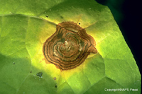 |
| Figure 6. Target spot lesion on tobacco caused by Rhizoctonia solani . (Courtesy H.D. Shew and T.A. Melton) |
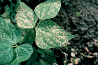
Bacterial leaf spots - spots are often angular due to limitation by leaf veins. Color is usually uniform and no signs of plant pathogen are evident. Tissue may appear initially as being water soaked but may become papery as it dries (Figure 7).
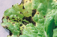 |
| Figure 7. Bacterial leaf spot on greenleaf lettuce caused by Xanthomonas campestris pv. vitians. (Courtesy S.T. Koike) |
Vein banding - Vein banding occurs when there is a band of yellow tissue along the larger veins of the leaf. This symptom is observed with viral diseases and is in contrast with nutrient deficiencies which may cause a dark green band along leaf veins (Figure 8).
 |
| Figure 8. Pea infected with Red clover vein mosaic virus exhibiting a vein chlorosis and banding. (Courtesy R. O. Hampton) |
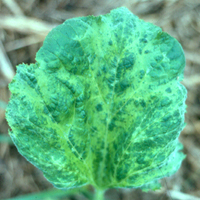
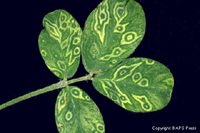
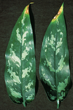
Mosaic and Ringspot - Mosaic (Figure 9) and ringspot (Figure 10) are used to describe an irregular patchwork of green and yellow areas over the surface of a leaf. In some cases leaves may also become distorted. Often these symptoms are associated with viral pathogens. There is not a sharp margin between the affected and healthy areas. Distinct margins may indicate a nutritional problem or genetic variegation.
 |
|
 |
|
| Figure 9. Mosaic symptoms exhibited on leaves of squash. (Used by permission of M. Riley, from files of W. Witcher) |
|
Figure 10. Peanut leaf with concentric ring spots caused by Tomato spotted wilt virus (TSWV). (Courtesy A. Culbreath, J. Todd, and H. Pilcher) |
|
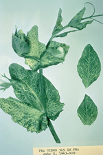
Leaf Distortion - Leaves of the infected plant may be distorted from their normal shape and size. Leaves may be elongated, smaller size, or thickened. This type of symptom can be associated with viral, fungal or bacterial infections (Figure 11) as well as insect and mite infestations.
 |
| Figure 11. Peach leaf curl caused by Taphrina deformans. (Courtesy J. W. Pscheidt) |
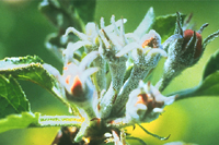
Powdery mildew - can affect leaves, stems, flowers and fruits with a white to gray surface coating of mycelia which can be rubbed off (Figure 5). Black specks may later develop in the mycelia. These specks are mature cleistothecia, the overwintering fungal structures which contain ascospores. Tissue may turn yellow, reddish or remain green under the mycelia and some leaf distortion may be observed especially on actively growing tissues.
|
| Figure 5. Powdery mildew on apple blossom cluster caused by Podospaera leucotricha . (Courtesy K.D. Hickey) |
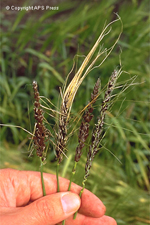
Presence of Spores/Spore Structures - Several fungal diseases can be easily identified based on the presence of spores or spore structures on the leaf surface. Some examples of this are rusts which are often recognized by the rusty brown to black spores (Figure 4) and smuts which are identified by the black spores which often replace the seed structure (Figure 12).
|
|
 |
|
| Figure 4. Stem rust on barley. caused by Puccinia graminis. (Courtesy B.Steffenson) |
|
Figure 12. Loose smut (on left) and false loose smut (on right) on barley, caused by Ustilago nuda (left) and U. nigra (right). (Courtesy P. Thomas) |
|
Needle drop in conifers - Conifers normally retain their needles for several years but these needles will eventually be lost. This drop is gradual and production of new needles obscures the loss of older needles. Unfavorable growing conditions, such as drought, may cause an acceleration of needle drop. If the drop occurs in only older needles especially during unfavorable growing conditions, there is no need for concern. If new needles are lost then other factors may be involved such as needle cast fungus (Figure 13), nutrient deficiencies, or toxic chemicals.
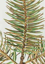 |
| Figure 13. Rhabdocline needle cast on Douglas Fir. (Courtesy E. Hansen) |
Chemical spray or air pollutant injury - Spots associated with injury are relatively uniform in color and the interface between the affected and healthy area is usually sharp. Distribution on plant may be associated with where spray or pollutant comes in contact with the plant. (Figure 14)
 |
| Figure 14. Injury of beans caused by drift of the herbicide, paraquat. (Courtesy H. F. Schwartz) |
Leaf or Needle Tip Death - Death at the tips of needles and tips and margins of leaves often indicate unfavorable climatic conditions, toxic chemicals or root malfunction due to poor cultural practices. Air pollutants, soil chemicals, and excess fertilizer can cause tip burn. Drought and freezing may have a similar effect. All needles of a specific growth period are usually affected. Needles infected by foliar fungal diseases are generally more scattered and rarely are all needles of a particular growth period killed (Figure 15). Needles diseased by infectious agents are generally affected over varying lengths and are often straw yellow or light tan. Fungal fruiting bodies may also be observed.
 |
| Figure 15. Chlorotic and necrotic lesions on leaf tips due to fluoride toxicity (Aglaonema commutatum 'Maria') (Courtesy J. M. F. Yuen) |
Soil or air chemical injury - Chemicals which are absorbed from the soil by roots or absorbed from air through leaves may exhibit a burning or scorching of leaf margins (Figure 16). If severe, islands of tissue between the veins may also be killed. Dead tissue may drop out of the leaf leaving a ragged appearance. Other chemicals may cause a distortion of leaf shape and size.
 |
| Figure 16. Alfalfa leaves exhibiting scorching of the leaf edges due to sulfur dioxide injury. (Courtesy K. T. Leath) |
Cankers - Cankers are localized necrotic lesions which are often sunken in appearance (Figure 17). Cankers can result from mechanical injury (e.g. trees which have been damaged by collisions with cars or lawnmowers), and various fungi or bacteria. In the spring, ooze may be observed on the surface of bacterial cankers and fruiting bodies may be observed on the surface of fungal cankers.
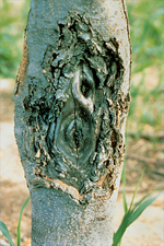 |
| Figure 17. Canker on apple caused by Nectria galligena. (Courtesy A. L. Jones) |
Fruit Decays and Rots - Various fungi and bacteria can cause rots of fruit. These are often distinguished by the color, lack of firmness of tissue, and signs of spores or fruiting bodies (Figure 18).
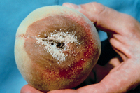 |
| Figure 18. Brown rot of peach caused by Monilinia fructicola. (Courtesy J. M. Ogawa) |
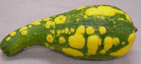
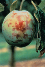
Fruit Discoloration - Discoloration of fruit is often associated with viral infections (Figure 19). This discoloration may be similar to mosaic and ringspot symptoms observed on leaves (Figure 20).
 |
|
 |
|
| Figure 19. Mosaic symptoms on yellow crookneck squash. Normal appearance is completely yellow fruit. (Used by permission of M. Williamson) |
|
Figure 20. Ringspot symptoms on peach fruit due to Plum pox virus. (Courtesy A. N. Adams) |
|
Wilts - Wilts are characterized by a general loss of turgidity of leaves or possibly entire plants due to the loss of water (Figure 21). The loss is most often caused by a blocking of the water flow through the xylem. This blockage can be caused by the presence of various bacteria (Erwinia, Ralstonia) and fungi (Fusarium, Verticillium) in the xylem. Wilts may also be observed when there is a destruction of the root system due to nematodes or root-rotting fungi (Armillaria, Phytophthora, Pythium) or an acute water shortage in the soil.
 |
| Figure 21. Verticillium wilt of cucumber caused by Verticillium dahliae. Courtesy W. D. Gubler) |
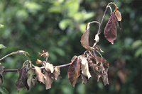
Shoot dieback or blights - Sudden dieback of a shoot usually indicates climatic or chemical injury rather than parasitic problems. If the line between affected and healthy bark is sharp, a soil chemical should be suspected. If dieback is somewhat more gradual and there is a cracking or splitting of the bark and wood, cold injury should be suspected along with bacterial blights caused by Pseudomonas or Erwinia. A bacterial streaming test with phase and compound microscope and isolation may be required to determine if the blights/dieback is due to a bacterial agent (Figure 3). Gradual decline of shoots and retention of dead leaves is more indicative of a parasitic disease (Figure 22). The margin between the affected and healthy tissue is often irregular and sunken. There may also be small pin-like projections or bumps over the surface of dead bark. These bumps are spore-producing structures of the fungal causal agent.
|
|
 |
|
| Figure 3. Bacterial ooze from cut tomato stem infected with Ralstonia solanacearum. (Used by permission of M. Williamson) |
|
Figure 22. Shoot growth of apple with fire blight, Erwinia amylovora, showing typical shepherd's-crook at the tip of the shoot. (Courtesy A. L. Jones) |
|
Dying branches of trees and shrubs - If scattered branches in a tree or shrub start to decline and eventually die, a canker disease or a shoot blight should be suspected (Figure 23). If branches die suddenly and, especially if affected branches are concentrated on one side of tree, weather conditions should be suspected (wind, snow, etc.) or animal or mechanical damage at the trunk base; however, this is not always the case. If symptoms develop over time on one side of a tree or plant then damage of the roots associated with one side may be suspected such as root rots due to Phytophthora spp.
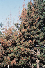 |
| Figure 23. Branch dieback of Monterey pine in California caused by pitch canker. (Courtesy L. D. Dwinell) |
Death of tree and shrub top - If all or a major portion of a tree or shrub dies over a period of time, the diagnostician should suspect a problem with the roots (Figure 24). Examples are diseases caused by Armillaria root rot and Verticillium wilt. The decline may be gradual and may eventually affect the entire tree, but in some cases the death may occur on one side of the plant initially. If the decline is sudden, a toxic chemical in soil or weather extremes such as freezing or drought should be suspected.
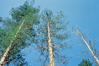 |
| Figure 24. Crown symptoms caused by laminated root rot of Douglas-fir. (Courtesy W. G. Thies) |
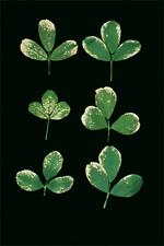
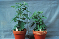
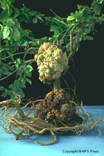
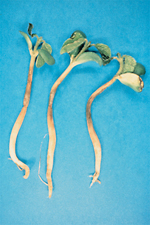
Overall Stunting or Decline - These symptoms can be caused by several very different factors. Systemic viral infections can result in stunting or decline, but such viral infections are often accompanied by other aboveground symptoms such as shortened internodes. In many cases, overall stunting of a plant may be due to problems associated with the root system (Figure 25). The roots should be examined for rotting and possible mycelial growth, reduction in roots especially feeder roots, and the presence of galls (Figure 26). Root galls can result from fungal and fungallike agents (Plasmodiophora brassicae), nematodes (Meloidogyne spp. - root-knot), and bacteria (Agrobacterium sp.). Abiotic factors such as nutritional deficiencies, soil compaction and herbicide residues can also result in overall stunting or decline.
 |
|
 |
|
| Figure 25. Healthy 'Mandalay' chrysanthemum (left) and plant infected with Fusarium oxysporum f. sp. chrysanthemi (right) which exhibits stunting without other observable symptoms. (Courtesy Penn State Univ.) |
|
Figure 26. Crown gall on Euonymous caused by Agrobacterium tumefaciens. (Courtesy Robert L. Forster) |
|
Damping-off - This term describes the rapid death and collapse of young seedlings. Often the seedlings will appear to be almost broken at the soil line (Figure 27). It may be observed in flats of plants begun in greenhouses and can result from infection of the seedlings by the fungal organisms Fusarium, Phytophthora, Pythium, Rhizoctonia, or Thielaviopsis (Figure 28).
 |
|
 |
|
| Figure 27. Soybean seedlings exhibiting symptoms of Pythium damping-off. Note thinning and browning of stem growth near the soil line. (Courtesy X. M. Yang) |
|
Figure 28. Damping-off of vinca (Catharanthus roseus) due to Rhizoctonia solani. (Courtesy R. L. Wick) |
|
Acknowledgment: Technical Contribution Number 4761 of the South Carolina Agricultural Experiment Station.
Citations
- Agrios, G. N. 1997. Introductory Plant Pathology. 4th ed. Academic Press, New York, NY.
- Alfieri, S. A., Jr., K. R. Langdon, J. W. Kimbrough, N. E. El-Gholl, and C. Wehlburg. 1994. Diseases and Disorders of Plants in Florida. Fla. Dep. Agric. Consumer. Serv. Div. Plant Ind. Bull. No. 14.
- Carlile, M. J., S. C. Watkinson, and G. W. Gooday. 2001. The Fungi, 2nd ed. Academic Press, New York, NY.
- Farr, D. F., G. F. Bills, G. P. Chamuris, and A. Y. Rossman. 1989. Fungi on Plants and Plant Products in the United States. American Phytopathological Society, St Paul, MN.
- Hansen, M. A. and R. L. Wick. 1993. Plant disease diagnosis: present and future prospects. Advances in Plant Pathology 10:65-126.
- Holmes, G. J., E. A. Brown, and G. Ruhl. 2000. What's a picture worth? The use of modern telecommunications in diagnosing plant diseases. Plant Dis. 84:1256-1265.
- Horst, R. K. 2001. Westcott's Plant Disease Handbook. 6th ed. Kluwer Academic Publishers, Boston, MA.
- Horst, R. K. 1983. Compendium of Rose Diseases. American Phytopathological Society, St Paul, MN.
- Kendrick, B. 2000. The Fifth Kingdom. 3rd edition. Focus Publishing, Newburyport, MA.
- Putnam, M. L. 1995. Evaluation of selected methods of plant disease diagnosis. Crop Protection 14:517-525.
- Shurtleff, M. C. and C. W. Averre. 1997. The plant disease clinic and field diagnosis of abiotic diseases. American Phytopathological Society, St. Paul, MN.
- United States Department of Agriculture. 1960. Index of Plant Diseases in the United States. Agricultural Handbook No. 165.
- Waller, J. M., B. J. Ritchie, and M. Holderness. 1998. Plant Clinic Handbook. CAB International, New York, NY.

